2022-05-13
10:15 - 11:15
Visual Computing Forum
Location
(II – 510N3 Blåbær (the ‘yoga’ room))
Speaker Biography
Heidi Espedal is a postdoctoral researcher at the Mohn Medical Imaging and Visualization Centre (MMIV) and the leader of the small-animal PET-CT facilities at the Molecular Imaging Center (MIC), University of Bergen. She is a medical cell biologist with a PhD in translational brain cancer research (2015) and 10 years of experience in preclinical magnetic resonance imaging (MRI) and positron emission tomography-computed tomography (PET-CT). Currently, her scientific focus is identifying early treatment response markers in endometrial cancer models.
Abstract
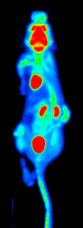 Imaging modalities such as magnetic resonance imaging (MRI) and positron emission tomography-computed tomography (PET-CT) is widely used for cancer diagnosis and management. In translational cancer research, animal models using patient-derived tumor and organoid xenograft models (PDX/O-PDX) represent useful tools for characterization of tumor growth and evaluation of new therapies. Preclinical imaging of PDX-models by MRI and PET-CT during disease progression enables visualization and quantification of functional tumor characteristics. The primary objective of this talk is to give an overview of current and novel preclinical multimodal imaging methods in cancer research.
Mailing List Subscription:
If you wish to receive email invitations to upcoming VCF talks/activities, please drop us an email (sherin.sugathan@uib.no) and we will gladly include your email in our mailing list.